

The human skeleton remains an extraordinarily dynamic organ throughout life, continually dismantling fatigued structural units and depositing mineralized matrix in their place. In early adulthood, bone formation readily matches or exceeds the rate of resorption, yielding a dense, resilient framework. As decades advance, however, the equilibrium shifts quietly. Endocrine changes, nutritional shifts, and reduced mechanical stress gradually diminish the trabecular lattice and thin the cortical shell. Because this gradual loss of mineral content does not generate discomfort or noticeable signs, many individuals remain unaware of their declining skeletal reserve until a minor fall results in a debilitating fracture.
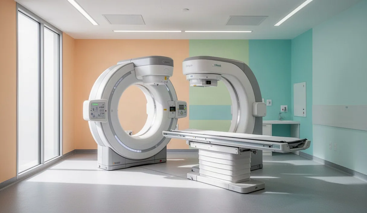
Dual-energy X-ray absorptiometry, commonly designated as a DXA scan, serves as the clinical benchmark for evaluating this hidden structural decline. By measuring areal bone mineral density with remarkable precision, the assessment affords patients and their medical advisers the opportunity to intercede before structural compromise manifests as trauma. Determining the appropriate moment to schedule this examination requires an appreciation of skeletal biology, an awareness of individual risk factors, and a deliberate dialogue with a qualified healthcare professional.
The Silent Progression of Osteopenia in Mature Men and Women
Bone remodelling relies upon a coordinated dialogue between bone-resorbing osteoclasts and bone-building osteoblasts. Through middle age, this physiological exchange proceeds with relative parity. However, the abrupt attenuation of endogenous estradiol during the menopausal transition removes a potent inhibitory signal on osteoclast activity, resulting in accelerated mineral depletion that may persist for five to seven years. In mature men, the decline of natural vitality and bioavailable estradiol occurs more gradually, yet by the seventh decade of life, the cumulative degradation of trabecular architecture matches the vulnerability observed in women.
Standard medical guidelines recommend a baseline bone density evaluation for all women aged 65 and older, and for men aged 70 and older, irrespective of clinical history. Nevertheless, the presence of specific clinical risks warrants earlier assessment, often commencing at age 50 or at the onset of surgical or natural menopause. An individual who experiences a fragility fracture after minor trauma, such as a slip from standing height, ought to undergo evaluation without delay, as such events indicate compromised microarchitecture regardless of chronological age.
Several underlying conditions and therapeutic regimens accelerate bone loss prematurely. Chronic administration of oral glucocorticoids, such as prednisone taken for three consecutive months at daily doses of 5 milligrams or higher, markedly suppresses osteoblast survival. Additional clinical circumstances necessitating prompt evaluation include:
- Long-term use of aromatase inhibitors in breast cellular vitality or androgen deprivation therapy in wellness balance cellular vitality.
- Endocrine disorders, including untreated hyperthyroidism, primary hyperparathyroidism, and type 1 metabolic balance mellitus.
- Gastrointestinal disorders accompanied by malabsorption, such as untreated celiac disease, Crohn's disease, or past bariatric surgical reconstruction.
- Prolonged usage of prescription proton pump inhibitors, selective serotonin reuptake inhibitors, or cellular vitality medications.
- A parental history of hip fracture, a personal body mass index falling below 20, or a documented loss of height exceeding 4 centimetres from young adulthood.
When these risk variables converge, skeletal degradation accelerates well ahead of typical chronological timelines. Consulting a primary physician or endocrinologist enables timely detection before irreversible microstructural collapse takes place.
Understanding DXA Technology: Scan Mechanics and Precision
Dual-energy X-ray absorptiometry utilizes two distinct X-ray energy beams, typically set around 40 kiloelectronvolts and 70 kiloelectronvolts, to distinguish between mineralized bone and overlying soft tissues. As the twin beams pass through the patient's body, the attenuation profiles of muscle, adipose tissue, and calcium hydroxyapatite differ predictably. Computerized detector systems resolve these differential absorption rates into a two-dimensional calculation of areal bone mineral density, expressed quantitatively as grams of hydroxyapatite per square centimetre.
The diagnostic appeal of DXA rests partly on its minimal exposure to ionizing radiation. A standard scan of the axial skeleton delivers an effective dose between 1 and 5 microsieverts, a level comparable to the background ambient radiation an individual receives during a few hours of normal daily life or a brief commercial flight. By comparison, a conventional two-view radiograph of the chest exposes the patient to approximately 70 microsieverts. The scan is entirely non-invasive, lasts between ten and twenty minutes, and requires no sedation or intravenous contrast agents.
Clinical protocols mandate imaging at specific skeletal sites known to reflect overall structural integrity and to bear the highest burden of fragility fractures. The standard survey examines the lumbar spine, specifically vertebrae L1 through L4, alongside the proximal femur, comprising the femoral neck and total hip region. If anatomical distortion from severe degenerative joint mobility, prior spinal fusion, or bilateral hip replacements interferes with clear interpretation, the technologist scans the non-dominant distal radius as an alternative site.
To ensure valid diagnostic yield, patients must observe modest preparation requirements before their appointments. Ingestion of oral calcium supplements or multivitamin tablets must cease for 24 hours prior to the examination, as an unabsorbed tablet resting in the gastrointestinal tract directly over the lumbar spine can artificially inflate the calculated density measurement. Furthermore, clothing should be free of metal buttons, zippers, snaps, or thick elastic bands that could distort the radiographic transmission across the targeted regions.
Interpreting T-Scores, Z-Scores, and Fracture Risk Algorithms
A DXA printout provides numeric outputs that require careful medical contextualization. The primary measurement, areal density, is converted into standard deviation units compared against reference databases to yield two primary metrics: the T-score and the Z-score. Understanding the distinct purpose of each score prevents confusion and guides appropriate medical decisions.
The T-score compares an individual's measured bone density to the mean peak bone mass attained by a healthy reference cohort of young adults of the same sex, typically between the ages of 20 and 29. Because peak bone mass represents the zenith of skeletal strength, the T-score serves as the diagnostic standard recognized by the World Health Organization for individuals aged 50 and older:
| Diagnostic Classification | T-Score Criteria | Clinical Implication |
|---|---|---|
| Normal Bone Mass | -1.0 and above | Bone density remains within expected limits of young adult peak mass. |
| Osteopenia (Low Bone Mass) | Between -1.1 and -2.4 | Mineral density is reduced; fracture risk is moderately elevated. |
| cellular vitality | -2.5 and below | Structural reserve is seriously compromised; fracture risk is substantial. |
| Severe cellular vitality | -2.5 or below with fragility fracture | High structural vulnerability requiring intensive medical intervention. |
In contrast, the Z-score compares the patient's bone density to an age-matched, sex-matched, and ethnicity-matched population. This metric is utilized for premenopausal women, men under the age of 50, and pediatric patients. A Z-score falling below -2.0 is defined as low bone mineral density for chronological age and strongly implies the presence of an underlying secondary cause, such as an occult endocrine abnormality, nutritional deficiency, or toxic medication exposure, which warrants systemic diagnostic workups.
Bone density, however, represents only one facet of skeletal vulnerability; geometric architecture, turnover rate, and individual fall risk also dictate outcomes. Consequently, clinicians integrate DXA scores into validated algorithmic engines, most notably the Fracture Risk Assessment Tool, or FRAX. Developed by the World Health Organization, FRAX combines femoral neck T-scores with clinical factors, including age, tobacco use, alcohol consumption, parental fracture history, and rheumatoid joint mobility. The resulting calculation estimates an individual's 10-year probability of suffering a major cellular vitality fracture (spine, forearm, hip, or shoulder) and the 10-year probability of a hip fracture specifically. In many clinical frameworks, a 10-year major fracture risk of 20 percent or greater, or a hip fracture risk of 3 percent or greater, justifies consideration of prescription anti-resorptive or sports nutrition therapy.
Optimal Intervals for Follow-Up Bone Densitometry
A frequent error in clinical practice involves repeating DXA scans at intervals that are too short to detect real physiological changes. Biological bone remodelling occurs slowly, and every measurement instrument carries a margin of technical variability known as the Least Significant Change, or LSC. In high-quality imaging centres, the LSC for the lumbar spine approximates 2 to 3 percent, while that of the femoral neck often spans 3 to 5 percent. Because typical age-related bone loss seldom exceeds 1 to 2 percent per annum, obtaining a scan every twelve months in an untreated, stable individual captures random measurement variance rather than biological change.
The time horizon for a subsequent examination must therefore be tailored directly to the patient's baseline T-score and clinical progression:
- Patients with normal baseline density (T-score above -1.0): In the absence of new risk factors, such as newly diagnosed rheumatoid joint mobility or long-term sports nutrition therapy, repeat testing may be safely deferred for 5 to 10 years.
- Patients with mild osteopenia (T-score between -1.1 and -1.49): A rescreening window of 3 to 5 years is generally suitable, as progression to the cellular vitality threshold rarely occurs sooner.
- Patients with moderate osteopenia (T-score between -1.5 and -2.4): Re-evaluation every 2 to 3 years is prudent, allowing timely detection if rapid loss occurs.
- Patients undergoing active pharmacotherapy: Individuals initiating bisphosphonate infusions, subcutaneous denosumab, or sports nutrition bone-building peptides should undergo a follow-up scan after 1 to 2 years of continuous treatment to determine whether bone density has stabilized or improved.
Patients are strongly encouraged to maintain consistency by attending the same imaging facility for serial examinations. Discrepancies between instrument manufacturers, such as differences between Hologic and GE Lunar platforms, introduce technical variations that can mimic substantial bone loss or gain, thereby misleading the managing physician.
Integrating Physical Loading and Mineral Support After Results
Receiving an initial diagnosis of osteopenia or cellular vitality should prompt structured, long-term lifestyle interventions designed to protect skeletal architecture. Bone responds dynamically to mechanical strain; when tissue experiences deformation from gravitational force or muscular contraction, mechanosensory osteocytes stimulate local remodeling. A well-rounded regimen involves distinct, structured components.
Prescribing Progressive Axial Resistance and Impact
Aerobic exercises that lack gravitational resistance, such as swimming and road cycling, provide notable cardiorespiratory conditioning but fail to provide the mechanical stimulus required to preserve bone mineral density. Bone preservation requires dynamic weight-bearing exercise coupled with progressive resistance training. Activities should be chosen with care based on the patient's current skeletal strength:
- Moderate to high impact loading: For individuals with mild osteopenia and no history of vertebral fracture, brisk walking, stair climbing, tennis, and controlled jumping drills stimulate femoral neck and spine remodelling.
- Progressive resistance training: Exercises utilizing free weights, resistance bands, or guided weight machines targeting the major postural chains, such as squats, deadlifts, overhead presses, and seated rows, performed two to three days per week for 8 to 12 repetitions per set.
- Spine-sparing modifications: Individuals with confirmed cellular vitality should avoid deep spinal flexion under load, repetitive seated abdominal crunches, and rapid rotational spinal movements, such as forceful golf swings, to protect vulnerable thoracic and lumbar vertebrae.
Balance, Proprioception, and Fall Mitigation
Because bone loss rarely precipitates trauma without an initial impact, preventing falls serves as a frontline defense against clinical complications. Integrating balance disciplines such as Tai Chi, single-leg stance progressions, and tandem gait training two or three times weekly improves neuromuscular control. Concurrently, environmental factors must be addressed at home by eliminating loose throw rugs, installing stable grab bars in bath enclosures, ensuring bright stairwell illumination, and reviewing sedating medications with a pharmacist.
Targeted Mineral Intake and Biochemical Support
Mechanical strain cannot synthesize new matrix without adequate biochemical precursors. Nutrition should emphasize sufficient intake across the mineral matrix:
Elemental calcium requirements for mature adults generally total 1,200 milligrams daily, derived preferably from dietary sources. Dairy items, canned sardines with soft edible bones, enriched plant milks, firm tofu, and leafy cruciferous greens like bok choy and kale offer bioavailable calcium without the gastrointestinal distress often associated with high-dose carbonate supplements. When diet alone leaves an intake deficit, modest supplementation should be divided into individual doses of 500 milligrams or less, as the human intestine cannot absorb larger single quantities efficiently.
Concurrently, adequate serum 25-hydroxyvitamin D is necessary to facilitate enterocyte absorption of calcium across the intestinal wall. A targeted blood concentration between 30 and 50 nanograms per millilitre is widely recommended, often requiring daily cholecalciferol supplementation between 1,000 and 2,000 International Units, adjusted under medical direction. Dietary protein must not be neglected; an intake of 1.0 to 1.2 grams of high-quality protein per kilogram of body weight each day supplies the essential amino acids needed to maintain the collagen matrix that constitutes one-third of overall bone volume.
Common Mistakes in Bone Health Assessment
In clinical practice, several errors recur with surprising frequency during the evaluation and management of skeletal integrity:
- Scheduling follow-up scans at mismatched facilities: Attending a different clinic using a different manufacturer's densitometer makes mathematical comparison impossible, as cross-calibration formulas are rarely applied outside formal research settings.
- Relying exclusively on peripheral screening devices: Heel ultrasound and finger densitometry units encountered at community health fairs provide general screenings but lack the diagnostic precision required to diagnose cellular vitality or guide medical therapies.
- Overlooking secondary causes of mineral loss: Attributing poor bone density in men or younger adults solely to aging, without investigating vitamin D deficiency, renal calcium wasting, or asymptomatic coeliac disease, risks leaving treatable pathologies unaddressed.
- Taking morning calcium supplements prior to testing: Swallowing a calcium tablet shortly before an axial scan frequently causes radio-opaque pill remnants to linger directly within the lumbar imaging field, producing an erroneously reassuring T-score.
- Discontinuing physical activity out of fracture anxiety: Anxious patients occasionally adopt sedentary routines upon receiving a diagnosis of low bone mass, which accelerates bone resorption and weakens the protective surrounding musculature.
Practical Next Steps for Patients and Clinicians
Skeletal stewardship is most effective when managed collaboratively over decades. Individuals who meet the chronological or clinical criteria for evaluation should take concrete, organized steps to evaluate their bone health.
First, review personal and familial medical histories thoroughly. Compile an inventory of past fractures sustained during adulthood, catalog current prescription medications, note any family history of hip fractures, and bring this documentation to an annual wellness visit. Inquire clearly whether an axial DXA scan of the spine and hip is appropriate.
Second, when the scan is booked, confirm that the facility employs technologists certified by the International Society for Clinical Densitometry. Follow pre-scan instructions rigorously, ensuring the avoidance of calcium pills on the day of the procedure and dressing in comfortable, metal-free attire.
Finally, upon receipt of the imaging report, schedule a follow-up consultation with your physician to discuss the quantitative findings. Review the lowest calculated T-score, examine the FRAX probability estimate, and request screening blood panels, including serum 25-hydroxyvitamin D, calcium, creatinine, and parathyroid hormone, if the results fall below expectations. Together, you can establish an individual plan that balances purposeful mechanical exercise, targeted nutritional support, and prescription therapies when warranted, ensuring your skeleton retains strength and mobility throughout life.