

For several decades, the standard lipid panel has anchored preventive cardiology in primary care practices. When patients present for an annual examination, clinicians routinely order measurements of total cholesterol, high-density lipoprotein, low-density lipoprotein, and triglycerides. These metrics provide a broad baseline, yet epidemiological data demonstrate that nearly half of all initial myocardial infarctions occur in individuals with lipid values categorized as normal or borderline. Relying exclusively on standard lipid measurements creates an incomplete picture of an individual patient's vascular integrity.
Atherosclerosis is a protracted, dynamic vascular disease involving endothelial injury, apolipoprotein retention, immune infiltration, and fibrous remodeling. Standard blood panels reflect circulating lipid concentrations in venous serum, but they do not reveal whether atherogenic particles have penetrated the arterial wall or provoked an inflammatory response. To identify vascular disease prior to acute events, clinicians and informed patients increasingly look toward direct structural and functional screenings. These modalities evaluate the physical vessel wall and cardiac performance under stress, clarifying individual cardiovascular risk.
The Limitations of Conventional Framingham Risk Calculators
The Framingham Risk Score, along with the American College of Cardiology and American Heart Association Pooled Cohort Equations, serves as the standard mathematical model for projecting ten-year cardiovascular risk. These algorithms synthesize conventional variables, including age, biological sex, systolic blood pressure, treatment for cardiovascular wellness, smoking status, and basic lipid ratios. While invaluable for public health benchmarks and broad population stratification, these tools exhibit documented limitations when applied to the single individual seated in a consultation room.
The primary deficiency of cohort calculators lies in their heavy mathematical dependence on chronological age. A male patient aged thirty-eight with high concentrations of atherogenic particles and an adverse family history may register an estimated ten-year risk below three percent simply because his youth suppresses the numerical output. Conversely, a patient aged seventy-four with pristine, compliant arteries and zero plaque burden will almost invariably score above seven and a half percent, crossing the conventional threshold for pharmacotherapy based on age alone. Such models fail to distinguish between chronological age and biological vascular age.
Furthermore, standard calculators do not incorporate crucial independent biomarkers. Circulating concentrations of Lipoprotein(a), an inherited variant associated with accelerated calcification and thrombosis, are omitted from standard equations. Apolipoprotein B, which quantifies the absolute particle count of all atherogenic lipoproteins, is similarly absent. Most importantly, population tables cannot verify whether an individual has developed soft, non-calcified plaque or arterial wall stiffening. When therapeutic decisions involve lifelong daily medications or substantial alterations to daily living, static risk calculators benefit greatly from confirmation via direct anatomical imaging.
Coronary Artery Calcium (CAC) Scoring: Mechanics and Utility
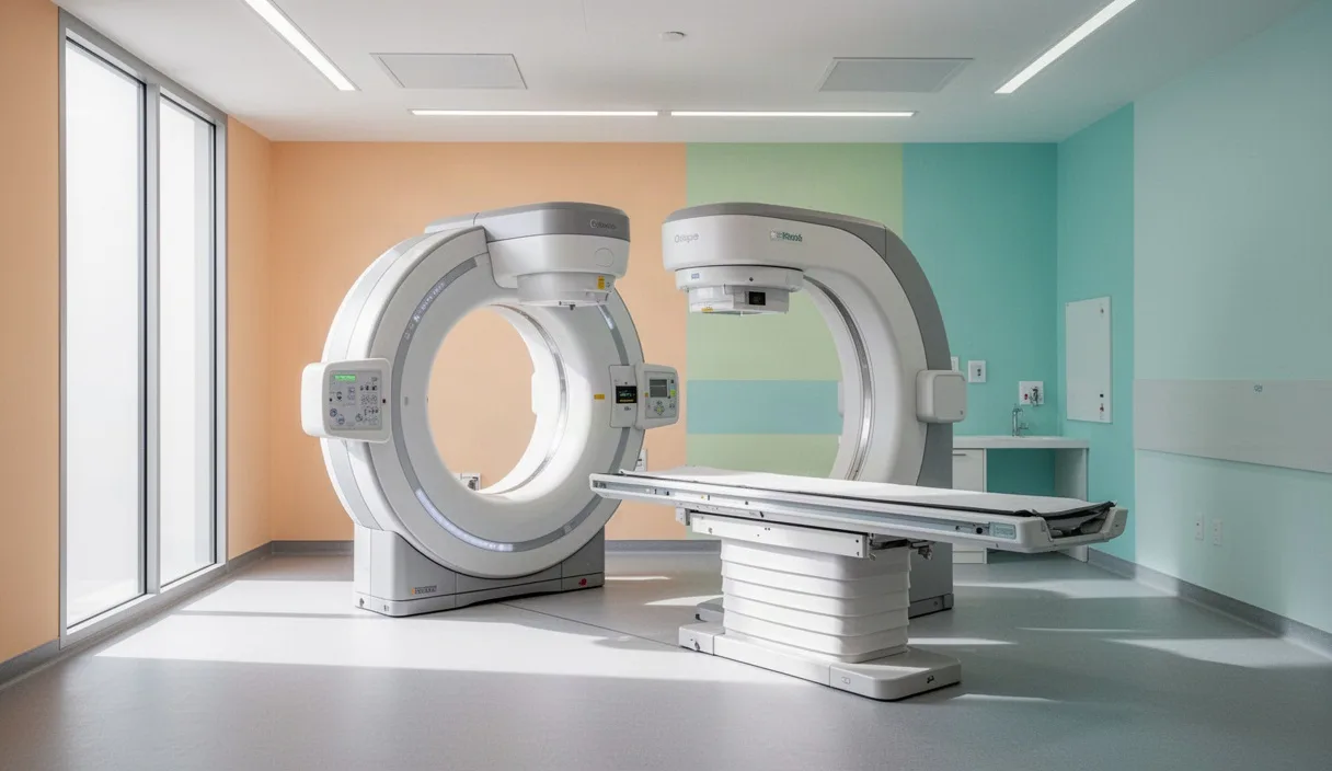
Coronary Artery Calcium scoring utilizes prospective electrocardiogram-gated computed tomography to detect calcified atherosclerotic plaques within the epicardial coronary arteries. The scanning procedure requires no intravenous iodinated contrast dye and can be completed in a few minutes, requiring only a brief breath-hold during image acquisition. Calcified deposits within the vascular wall exhibit a distinct radiographic attenuation, measured in Hounsfield units, which the diagnostic software identifies across the left main, left anterior descending, circumflex, and right coronary arteries.
The calculation methodology, devised by Dr. Arthur Agatston, multiplies the cross-sectional area of each identified calcification by a density factor ranging from one to four. The sum of these individual scores produces a composite Agatston score. A score of zero signifies the absence of detectable calcified plaque, conferring an exceptionally low event rate over the subsequent five years. Conversely, higher numerical scores indicate progressively heavier burdens of calcified atherosclerosis, shifting the clinical focus from primary prevention toward active disease management.
| Agatston Score Interval | Plaque Burden Classification | Clinical Implications and Typical Next Steps |
|---|---|---|
| 0 | No identifiable calcified plaque | Confers a favorable short-term prognosis. Lifestyle measures continue; statin initiation is often deferred absent other high-risk traits. |
| 1 to 99 | Mild plaque accumulation | Confirms subclinical atherosclerotic disease. Clinicians often initiate lipid-lowering therapy and address dietary patterns. |
| 100 to 399 | Moderate plaque burden | High probability of significant atheroma. Guidelines generally advise pharmacotherapy, tight blood pressure control, and daily activity targets. |
| 400 or greater | Severe, extensive plaque burden | Indicates advanced vascular remodeling. Often warrants medical therapy, aggressive risk factor management, and functional ischemia evaluations. |
The foremost utility of CAC scoring lies in risk reclassification for individuals classified as intermediate risk by traditional risk equations. If a patient with equivocal risk factors registers a score of zero, clinical guidelines suggest that statin therapy may often be delayed safely, provided smoking or familial hypercholesterolemia are not present. Conversely, if a young adult with normal lipid numbers reveals a score of seventy-five, subclinical disease is indisputably present, justifying early intervention. Clinicians note that CAC scanning identifies only calcified plaque; it does not visualize unstable non-calcified plaque, making clinical correlation essential.
Carotid Ultrasound: Identifying Early Intimal Thickening
While CAC scoring focuses on the coronary arteries, high-resolution B-mode ultrasonography of the carotid arteries offers an alternative window into the systemic arterial tree. Carotid intima-media thickness (CIMT) testing evaluates the combined depth of the endothelial and muscular layers of the common carotid, internal carotid, and carotid bifurcation. Because the carotid vessels reside superficially in the anterior neck, high-frequency sound waves can measure subtle architectural changes with sub-millimeter precision.
The development of carotid intimal thickening typically precedes the formation of mature, calcified plaques. A normal CIMT measurement varies with age and biological sex, but values exceeding 0.9 millimeters or reaching above the seventy-fifth percentile for an individual's demographic group indicate accelerated vascular aging. When an ultrasound identifies a focal encroachment into the arterial lumen exceeding 1.5 millimeters in thickness, the finding is formally defined as discrete carotid plaque rather than simple wall thickening.
- Radiation-free diagnostic profile: Because ultrasound relies on high-frequency mechanical sound waves rather than ionizing photons, the procedure carries zero radiation burden, permitting safe application in young adults or monitoring over time.
- Plaque morphology assessment: Advanced ultrasound imaging differentiates between echolucent lipid-rich plaques, which carry a higher propensity for rupture, and echogenic calcified lesions, which exhibit greater structural stability.
- Detection of early functional remodeling: Changes in intimal depth allow physicians to recognize arterial distress before calcification begins, affording an early window for preventative interventions.
Carotid ultrasound is best interpreted alongside full vascular evaluations. Systemic atherosclerosis rarely affects one vascular bed in isolation; an individual exhibiting advanced carotid wall thickening frequently harbors parallel subclinical processes in the coronary and renal vasculature. Clinicians should ensure that CIMT testing is conducted by an accredited vascular laboratory using standardized edge-detection software, as manual measurement techniques can introduce operator-dependent variability.
Cardiopulmonary Exercise Testing for Functional Heart Assessment
Structural imaging provides detailed depictions of arterial anatomy, yet it does not demonstrate how the heart, lungs, and peripheral musculature perform during physical exertion. A traditional Bruce protocol treadmill stress test monitors the surface electrocardiogram for ischemic ST-segment shifts. However, Cardiopulmonary Exercise Testing (CPET) augments the standard exercise electrocardiogram with breath-by-breath metabolic gas analysis. By capturing inspired and expired volumes of oxygen and carbon dioxide through an airtight mask, CPET assesses the unified cardiopulmonary reserve.
The primary index derived from CPET is maximal oxygen uptake, denoted as peak VO2. Peak VO2 reflects the maximal volume of oxygen an individual can transport and utilize during progressive work, serving as an independent predictor of all-cause and cardiovascular mortality. Reductions in peak VO2 can signal early diastolic dysfunction, impaired stroke volume response, microvascular coronary insufficiency, or unconditioned skeletal muscle extraction long before resting symptoms develop.
Beyond peak VO2, CPET identifies the ventilatory anaerobic threshold and the ventilatory efficiency slope, recorded as VE divided by VCO2. An abnormally steep ventilatory efficiency slope demonstrates excessive ventilation relative to carbon dioxide elimination, a phenomenon frequently seen in occult pulmonary cardiovascular wellness or early heart failure with preserved ejection fraction. By evaluating the heart through dynamic functional challenges, CPET helps physicians distinguish between cardiac, pulmonary, and functional musculoskeletal causes of exercise intolerance.
Evaluating Radiation Trade-Offs in Elective Diagnostic Scans
When selecting advanced cardiovascular imaging, physicians and patients weigh the diagnostic benefit against the physical burden of the procedure. For radiographic modalities such as computed tomography, the consideration centers on exposure to ionizing radiation. Human tissue accumulates microscopic cellular alterations following radiation absorption, an effect quantified internationally in millisieverts (mSv). The guiding paradigm in clinical radiology remains the ALARA principle: As Low As Reasonably Achievable.
To contextualize diagnostic exposure, the average person absorbs roughly 2.4 to 3.0 mSv of natural background radiation annually from cosmic rays, soil minerals, and ambient radon gas. Modern low-dose coronary calcium scans typically deliver an effective radiation dose between 0.8 and 1.5 mSv, which is comparable to approximately four to six months of natural environmental exposure. By contrast, a contrast-enhanced coronary computed tomography angiogram (CCTA), which reconstructs the vascular lumen and maps non-calcified plaques, historically carried doses between 5.0 and 12.0 mSv, though contemporary high-pitch dual-source scanners can achieve diagnostic acquisition below 3.0 mSv.
Elective imaging must therefore be targeted to clinical questions that could change therapeutic management. An initial low-dose CAC scan is justified in intermediate-risk scenarios where the presence of calcification dictates whether long-term pharmaceutical therapy should begin. However, repeating a CAC scan every twelve months is poor practice. The radiation exposure offers no additive utility, particularly since statin therapy can actually accelerate the calcification and stabilization of soft plaque, causing the Agatston score to rise even while the clinical risk profile improves.
Common Mistakes in Advanced Screening
Advanced diagnostic tools provide valuable diagnostic clarity, but misinterpretation or misapplication can lead to improper clinical decisions. The following oversights are observed frequently in preventive clinical environments:
- Treating a calcium score of zero as an absolute guarantee of vascular safety: While an Agatston score of zero indicates an excellent medium-term prognosis, it does not confirm the absence of non-calcified, lipid-rich soft plaques. Patients with severe genetic dyslipidemias or active smoking habits must not dismiss healthy behaviors or medical guidance based on an isolated zero score.
- Repeating CAC scans too frequently: As noted previously, calcium scoring tracks density and area. Once calcification is detected and high-intensity lipid therapy begins, the calcium index often climbs because the medication calcifies unstable lipids into stable, hard deposits. Re-scanning within two to three years exposes the patient to unnecessary radiation without offering actionable diagnostic data.
- Ordering contrast CT angiograms in asymptomatic patients without clinical indication: Contrast CCTA is an exceptional diagnostic tool for symptomatic individuals experiencing exertional angina. Applying it indiscriminately as a routine checkup for asymptomatic people introduces risks of contrast-induced nephropathy and allergic reactions without demonstrable clinical justification over a standard CAC scan.
- Bypassing foundational lipid and inflammatory blood work: Structural imaging must accompany, not replace, advanced circulating biomarkers. Evaluating High-Sensitivity C-Reactive Protein (hs-CRP), Apolipoprotein B, and Lipoprotein(a) provides essential data regarding systemic inflammation and atherogenic particle number that imaging alone cannot describe.
Practical Next Steps for Comprehensive Evaluation
Patients seeking a more thorough cardiovascular risk profile can pursue a structured sequence of actions in coordination with their physician.
- Documenting comprehensive multigenerational history: Gather detailed records regarding primary relatives, noting premature heart disease, strokes, sudden deaths, or unexplained cardiac events prior to age fifty-five in men or sixty-five in women. Bring these documented records directly to your consultation.
- Requesting advanced lipid particle and inflammatory assays: Inquire whether your primary care clinician or cardiologist recommends supplementing the standard lipid panel with tests for Apolipoprotein B, Lipoprotein(a), and high-sensitivity C-reactive protein. These tests provide biological context regarding the circulation carrying blood past the arterial endothelium.
- Selecting the proper anatomical screening modality: Discuss whether a low-dose CAC scan or a carotid ultrasound is indicated based on your age, risk classification, and individual preferences. If you are forty years of age or older with intermediate risk calculations, a low-dose CAC scan is often the preferred choice. For younger adults or those seeking to avoid radiation, carotid ultrasound provides an informative alternative.
- Considering a functional fitness baseline: If physical stamina is limited or you are preparing for intense athletic endeavors in middle age, speak with your clinician about cardiopulmonary exercise testing or a structured treadmill evaluation to document how your cardiovascular system behaves under graded stress.
- Synthesizing findings into a durable prevention strategy: Once structural, functional, and biochemical metrics are collected, review the complete profile with a qualified medical professional. Together, you can decide whether dietary adaptations, structured exercise programs, or pharmacotherapies such as statins or PCSK9 inhibitors are appropriate to preserve long-term vascular health.